논문리뷰 : 요통을 느끼지 않는 사람들의 요추 MRI 자기공명영상 : Published July 14, 1994 N Engl J Med 1994;331:69-73
작성자 정보
- 삼둡 작성
- 작성일
컨텐츠 정보
- 7,776 조회
-
목록
본문

https://www.nejm.org/doi/full/10.1056/NEJM199407143310201
AbstractBackground
The relation between abnormalities in the lumbar spine and low back pain is controversial. We examined the prevalence of abnormal findings on magnetic resonance imaging (MRI) scans of the lumbar spine in people without back pain.
Methods
We performed MRI examinations on 98 asymptomatic people. The scans were read independently by two neuroradiologists who did not know the clinical status of the subjects. To reduce the possibility of bias in interpreting the studies, abnormal MRI scans from 27 people with back pain were mixed randomly with the scans from the asymptomatic people. We used the following standardized terms to classify the five intervertebral disks in the lumbosacral spine: normal, bulge (circumferential symmetric extension of the disk beyond the interspace), protrusion (focal or asymmetric extension of the disk beyond the interspace), and extrusion (more extreme extension of the disk beyond the interspace). Nonintervertebral disk abnormalities, such as facet arthropathy, were also documented.
Results
Thirty-six percent of the 98 asymptomatic subjects had normal disks at all levels. With the results of the two readings averaged, 52 percent of the subjects had a bulge at at least one level, 27 percent had a protrusion, and 1 percent had an extrusion. Thirty-eight percent had an abnormality of more than one intervertebral disk. The prevalence of bulges, but not of protrusions, increased with age. The most common nonintervertebral disk abnormalities were Schmorl's nodes (herniation of the disk into the vertebral-body end plate), found in 19 percent of the subjects; annular defects (disruption of the outer fibrous ring of the disk), in 14 percent; and facet arthropathy (degenerative disease of the posterior articular processes of the vertebrae), in 8 percent. The findings were similar in men and women.
Conclusions
On MRI examination of the lumbar spine, many people without back pain have disk bulges or protrusions but not extrusions. Given the high prevalence of these findings and of back pain, the discovery by MRI of bulges or protrusions in people with low back pain may frequently be coincidental.
배경
요추의 이상과 요통 사이의 관계는 논란의 여지가 있습니다. 우리는 요통이 없는 사람들의 요추 자기공명영상(MRI) 스캔에서 이상 소견의 유병률을 조사했습니다.
조사 방법
무증상자 98명을 대상으로 MRI 검사를 실시했습니다. 피험자의 임상 상태를 모르는 두 명의 신경 방사선 전문의가 독립적으로 스캔을 판독했습니다. 연구 해석의 편향 가능성을 줄이기 위해 요통이 있는 27명의 비정상적인 MRI 스캔을 무증상자의 스캔과 무작위로 혼합했습니다. 요추의 추간판 5개를 정상, 팽륜(bulge, 디스크가 간격 너머로 원주 대칭적으로 확장), 돌출(protrusion, 디스크가 간격 너머로 국소 또는 비대칭적으로 확장), 탈출(디스크가 간격 너머로 더 극단적으로 확장) 등의 표준화된 용어를 사용하여 분류했습니다. 파셋 관절증(facet arthropathy)과 같은 비추간판 이상도 문서화되었습니다.
결과
무증상 피험자 98명 중 36%는 모든 레벨에서 디스크가 정상으로 나타났습니다. 두 번의 측정 결과를 평균한 결과, 피험자의 52%는 적어도 한 단계 이상에서 팽륜이 있었고 27%는 돌출이 있었으며 1%는 탈출이 있었습니다. 38%는 두 개 이상의 추간판에 이상이 있었습니다. 돌출은 아니지만 팽륜된 추간판의 유병률은 나이가 들수록 증가했습니다. 가장 흔한 비추간판 이상은 슈몰 노드(Schmol's nodes디스크가 척추체 끝판으로 탈출하는 질환)로 전체 대상자의 19%에서 발견되었으며, 환형 결손(annual defects,디스크 외부 섬유륜의 파괴)은 14%, 척추 관절증(facet arthropahy, 척추의 후관절 돌기의 퇴행성 질환)은 8%에서 발견되었습니다. 이러한 결과는 남성과 여성에서 비슷하게 나타났습니다.
결론
요추 MRI 검사에서 요통이 없는 많은 사람들은 디스크가 튀어나오거나 돌출되어 있지만 추간판 탈출증은 없습니다. 이러한 소견과 요통의 높은 유병률을 고려할 때 요통 환자의 디스크 팽륜 또는 돌출이 MRI에서 발견되는 것은 종종 우연일 수 있습니다.

표 1은 MRI 검사에서 **디스크 팽륜 (bulges), 돌출(protrusions), 탈출(extrusions)**의 유병률을 요약한 것입니다.
두 명의 판독자 결과를 평균한 결과, 무증상자(허리 통증이 없는 사람)의 52%가 한 개 이상의 추간판에서 팽륜이 있었고, 27%가 돌출, 1%가 탈출이 있었습니다. 즉, 허리 통증이 전혀 없는 사람들 중 64%가 디스크 이상 소견을 보였고, 38%는 두 개 이상의 부위에서 이상이 발견되었습니다.

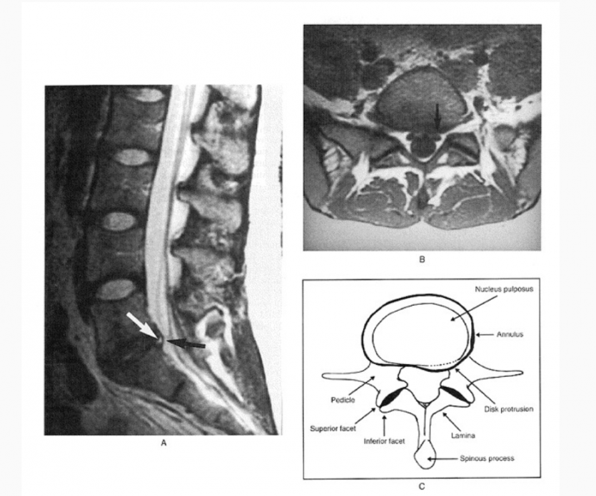
허리 통증이 없는 24세 여성의 디스크 돌출 MRI와 그 도식적 표현입니다.
성별과 디스크 팽륜 유병률 사이에는 유의미한 관련성이 없었고, 연령과 돌출 유병률 사이에도 유의미한 관련성은 없었습니다.
디스크 팽륜은 나이가 들수록 유병률이 증가했으며(P < 0.001), 이 경향은 모든 추간판 레벨에서 나타났습니다.
무증상자에서 발견된 가장 흔한 디스크 외 이상 소견
슈몰 결절(Schmorl’s nodes) (디스크가 척추체 연골판 안으로 탈출된 상태): 19%
섬유륜 결손(Annular defects) (디스크 바깥 섬유 고리의 손상): 14%
관절돌기병(Facet arthropathy) (척추 후방 관절의 퇴행성 병변): 8%
척추 분리증(Spondylolysis): 7%
척추 전방전위증(Spondylolisthesis): 7%
척추 중앙관 협착(Central canal stenosis): 7%
신경공 협착(Neural foramen stenosis): 7%
관련자료
-
이전
-
다음


