c1의 촉진 ; palpation of c1 vertebra
작성자 정보
- 삼둡 작성
- 작성일
컨텐츠 정보
- 11,212 조회
- 목록
본문
Validity of palpation of the C1 transverse process: comparison with a radiographic reference standard
Robert Cooperstein, MA, DC,a,b Morgan Young, DC,a,b and Makani Lew, DCa
Author information Copyright and License information PMC Disclaimer
Abstract
Objectives:
Primary goal: to determine the validity of C1 transverse process (TVP) palpation compared to an imaging reference standard.
Methods:
Radiopaque markers were affixed to the skin at the putative location of the C1 TVPs in 21 participants receiving APOM radiographs. The radiographic vertical distances from the marker to the C1 TVP, mastoid process, and C2 TVP were evaluated to determine palpatory accuracy.
Results:
Interexaminer agreement for radiometric analysis was “excellent.” Stringent accuracy (marker placed ±4mm from the most lateral projection of the C1 TVP) = 57.1%; expansive accuracy (marker placed closer to contiguous structures) = 90.5%. Mean Absolute Deviation (MAD) = 4.34 (3.65, 5.03) mm; root-mean-squared error = 5.40mm.
Conclusions:
Manual palpation of the C1 TVP can be very accurate and likely to direct a manual therapist or other health professional to the intended diagnostic or therapeutic target. This work is relevant to manual therapists, anesthetists, surgeons, and other health professionals.
Keywords: palpation, C1 TVP, radiograph, validity, chiropractic
C1 횡돌기 촉진의 유효성: 방사선 참조 표준과의 비교
로버트 쿠퍼스타인, MA, DC,a,b 모건 영, DC,a,b 및 마카니 루, DCa
저자 정보 저작권 및 라이선스 정보 PMC 면책 조항
바로가기:
초록
목적:
일차 목표: 영상 참조 표준과 비교하여 C1 횡돌기(TVP) 촉진의 유효성을 확인합니다.
방법:
APOM 방사선 사진을 촬영한 21명의 참가자에게 C1 TVP 추정 위치의 피부에 방사선 불투명 마커를 부착했습니다. 마커에서 C1 TVP, 유양돌기 및 C2 TVP까지의 방사선 사진 수직 거리를 평가하여 촉진 정확도를 결정했습니다.
결과:
방사선 측정 분석에 대한 검사자 간 일치도는 "우수"였습니다. 엄격 정확도(C1 TVP의 가장 외측 돌출부에서 ±4mm 떨어진 곳에 마커 배치) = 57.1%, 확장 정확도(인접한 구조에 더 가깝게 마커 배치) = 90.5%. 평균 절대 편차(MAD) = 4.34(3.65, 5.03) mm; 제곱근 평균 오차 = 5.40mm.
결론:
C1 TVP의 수동 촉진은 매우 정확할 수 있으며 도수 치료사 또는 기타 의료 전문가가 의도한 진단 또는 치료 목표에 도달할 수 있도록 도와줄 가능성이 높습니다. 이 작업은 도수 치료사, 마취과 의사, 외과 의사 및 기타 의료 전문가와 관련이 있습니다.
키워드: 촉진, C1 TVP, 방사선 사진, 유효성, 카이로프랙틱

True horizontal lines A, B, C, and D demarcate the inferior mastoid, C1 TVP, marker, and C2 TVP, respectively. The red vertical lines pass through the markers. Line B-C is examiner error for C1 TVP palpation, line A-C is maker-mastoid distance, and line D-C is C2 TVP–marker distance. In this exemplar radiograph, the right mastoid process and the upper surface of the left C1 TVP are difficult to visualize.
실제 수평선 A, B, C, D는 각각 하비갑개, C1 TVP, 마커 및 C2 TVP를 구분합니다. 빨간색 수직선은 마커를 통과합니다. 선 B-C는 C1 TVP 촉진에 대한 검사자 오차, 선 A-C는 제작자-유양돌기 거리, 선 D-C는 C2 TVP-마커 거리입니다. 이 예시 방사선 사진에서 오른쪽 유양돌기 과정과 왼쪽 C1 TVP의 상부 표면은 시각화하기 어렵습니다.
In the present study, accuracy was calculated in both a stringent and a more permissive manner. According to the strict definition, a palpator would place a soft-tissue marker within the 8mm wide field of the C1 TVP; whereas according to the more clinically relevant expansive definition, the palpator would place the marker closer to the C1 TVP than to either the mastoid process above or the C2 TVP below. Unlike all but one of the spinal landmark validity studies reviewed, our study involved TVP rather than SP palpation. The only other study to have done so was Jende et al14, who found palpation to have been inaccurate. Due to factors related to image interpretation, it would have been difficult to define a field of marker accuracy by projecting lines from the inferior and superior aspects of TVPs, analogous to what investigators had done in the reviewed SP studies. As an alternative method, the authors identified the most lateral projection of the C1 TVP on the radiograph, which moreover would have presumably been the most likely aspect of the vertebra with which the palpator had “made contact” through the soft tissue.
본 연구에서는 정확도를 엄격한 방식과 보다 허용적인 방식으로 계산했습니다. 엄격한 정의에 따르면 촉진자는 C1 TVP의 8mm 너비 범위 내에 연부조직 마커를 배치하는 반면, 임상적으로 보다 포괄적인 정의에 따르면 촉진자는 마커를 위쪽 유양돌기나 아래쪽 C2 TVP보다 C1 TVP에 더 가깝게 배치합니다. 검토된 척추 랜드마크 유효성 연구 중 한 가지를 제외한 모든 연구와 달리, 본 연구에서는 SP 촉진이 아닌 TVP를 사용했습니다. 그렇게 한 유일한 다른 연구는 Jende 등14 으로, 촉진이 정확하지 않다는 것을 발견했습니다. 이미지 해석과 관련된 요인으로 인해 검토된 SP 연구에서 연구자들이 수행한 것과 유사하게 TVP의 열등한 측면과 우수한 측면에서 선을 투영하여 마커 정확도 영역을 정의하는 것은 어려웠을 것입니다. 이에 대한 대안으로 저자들은 방사선 사진에서 C1 TVP의 가장 측면에 투영된 부분을 확인했으며, 이는 아마도 촉진기가 연부 조직을 통해 "접촉"한 척추의 가장 가능성이 높은 측면이었을 것입니다.
Our method of calculating expanded accuracy, wherein the marker was closer to the target than to contiguous structures, necessarily differed in method from some of the SP validity studies that also included a concept of liberal accuracy.4,11,13,41,42 In these other studies the contiguous structures were equally distant from the targeted structure; whereas in the present study, the mastoid-C1 TVP distance did not equal to the C1 to C2 TVP distance. Since the average distance of marker to mastoid was 8.70mm, and to C2 TVP midpoint 13.77mm, there was no way to define a constant magnitude of examiner error that would be regarded as expansively accurate. Therefore, the authors defined expanded accuracy in a purely clinical sense as having placed the center of the radiopaque marker closer to either of the contiguous structures (mastoid process and C2 TVP).
마커가 인접 구조물보다 표적에 더 가까운 확장 정확도를 계산하는 본 연구의 방법은 자유 정확도 개념을 포함하는 일부 SP 타당도 연구와 방법이 다를 수밖에 없었습니다.4,11,13,41,42 이러한 다른 연구에서는 인접 구조물이 표적 구조물에서 똑같이 멀리 떨어져 있었지만, 본 연구에서는 유양돌기-C1 TVP 거리가 C1과 C2 TVP 거리와 같지 않았습니다. 마커에서 유양돌기까지의 평균 거리가 8.70mm, C2 TVP 중간점까지의 거리가 13.77mm였기 때문에, 확장된 정확도로 간주할 수 있는 검사자 오차의 일정한 크기를 정의할 방법이 없었습니다. 따라서 저자들은 순전히 임상적 의미에서 확장된 정확도를 방사선 불투명 마커의 중심을 인접한 구조물(유양돌기 및 C2 TVP) 중 하나에 더 가깝게 배치한 것으로 정의했습니다.
0:00
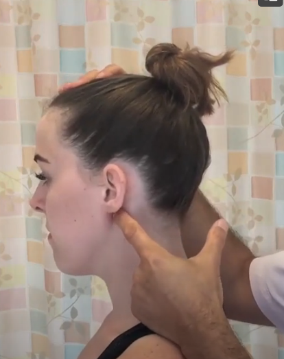
오늘은 C1을 촉진해 보겠습니다.
0:03
횡단 과정과 첫 번째 경로
0:05
그래서 C1 가로 프로세스의 경우
0:08
에 가장 먼저 할 일은
0:09
유양 돌기 과정 그리고 당신은 할 것입니다
0:11
턱의 선을 따라 약
0:13
반쯤 내려 가면 가로가 느껴집니다.
0:19
C1의 과정과 당신이하는 방식
0:21
이것이 횡단임을 알고
0:23
C1의 프로세스는 여러분이
0:25
머리를 앞뒤로 움직여 하나를 볼 수 있습니다.
0:28
의 회전
경추부 극돌기의 해부학적 표식점은 경추에서 발생 하는 통증의 정확한 위치를 확립하는데 있어 매우 중요 하다고 한다(척추정형내과연구회, 1999)[그림 1]. 그림 1. 경추의 표식점 C6, C5, C4, 그리고 C3의 극돌기들은 거의 같은 정 도로 뒤쪽으로 돌출되어 있다. 그러나, C2 극돌기는 다 른 경추 극돌기 보다 더 크고, 더 돌출되어 있으며, 귓 불(ear lobe)레벨 아래쪽에 위치하고 있다고 하며(채윤 원 , 2008), 이현옥 등 (2008)은 외후두 융기를 확인하 고, 그대로 C7극돌기를 향하여 가볍게 압박하면서 손 가락을 진행 시킨다. 최초로 느끼는 뼈 돌출부가 C2극 돌기 이다(대한정형도수치료학회, 2004. ; 척추정형내 과연구회 ,1999. ; 정진우, 1986.) 또 다른 방법으로 후두골(occipital bone) 아래 부분 에 작은 함몰부가 있으며, 함몰부 아래쪽에서 시술자는 골성구조를 감지할 수 있을 것이다. 이것이 C2(축추)의 극돌기이다. C2 극돌기는 상당히 튀어나와 있으며, 대 상자의 이마에 손을 올리고 부드럽게 대상자의 머리를 약간 골곡시키고 신전시킴으로 분명하게 확인할 수 있 다고 하였다(Cyriax 정형의학연구회 ,2009
It is not uncommon for this to occur. It is possibly an asymmetry in the bone, but more likely a static malposition of the atlas. Whether it matters functionally would have to be determined on a case by case basis.
이런 일이 발생하는 것은 드문 일이 아닙니다. 뼈의 비대칭일 수도 있지만, 아틀라스의 정적 위치가 잘못되었을 가능성이 더 높습니다. 기능적으로 중요한지 여부는 사례별로 판단해야 합니다.
관련자료
-
이전
-
다음


